Necker Hospital Pediatric Dermatology Seminar (SDPHN)
Held last June at the Maison de la Chimie in Paris, this seminar is a great success every year, bringing together mainly pediatricians and dermatologists. Presentations on the latest developments are given in the mornings, while the afternoons are devoted to illustrated clinical cases.

Here, we summarize the presentations on the latest developments in contact eczema in children, the diagnosis of phytophotodermatoses and the problems posed by pediatric rashes.
1. Contact dermatitis in children: frequent and under-diagnosed
Contact dermatitis (CD) is common in children (16.5% in the United States), but largely under-diagnosed. It is responsible for 20% of atopic dermatitis (AD). Contact sensitization is found in 40%-50% of children tested after referral.
EC can be observed from the very first months of life, and its frequency increases with age.
Risk factors for sensitization to topicals in AD are the severity and precocity of AD, and IgE-mediated sensitization.
CE should be considered in the presence of certain suggestive lesion distributions (palmoplantar, perioral, periumbilical and sub-elastic zones, seat). A suggestive clinical history, activities and hobbies such as modelling clay or painting, a dermatosis of unknown origin, a worsening of a previously stable dermatosis and the absence of improvement under treatment of an eczema should also prompt a search for CE.

Clinical presentations of CE
- Extensive erythematous maculopapular lesions
- Embedded palmar and digital vesicles
- Erythema of extremity folds
- Confluent erythema of perineal area and folds
- Perioral or labial dermatitis, cheilitis
- Eyelid dermatitis or edema
To identify the allergens involved, the current European standard battery includes twenty-six allergens most frequently responsible for allergic contact dermatitis. For children, the standard battery will be simplified, prioritizing the allergens most frequently found in children (metals, perfumes, surfactants, local antibiotics, preservatives) and adding personal products. Targeted questioning according to age is therefore essential.
Contact allergies to antiseptics
Contact eczemas to antiseptics, which are rarely reported in children, are under-diagnosed. They require patch-testing or, at the very least, an open test of repeated application (ROAT test) with the suspected product, to characterize the allergen.
Chlorhexidine digluconate is the predominant allergen in contact eczema to antiseptics in children. Co-sensitization is frequent, notably with benzalkonium chloride and benzyl alcohol, sometimes present in the same preparation. Hexamidine (family of biguanides and amidines) may also be involved. The question is whether biguanides should continue to be widely used as disinfectants, and whether they should be banned in atopic subjects.
How can contact dermatitis be investigated?
Patch tests are used to investigate delayed allergies mediated by T lymphocytes. A standard battery is used, with certain specificities (addition of personal products).
ROAT tests, like use tests and evictions, are useful alternatives to patch tests.
Management of contact dermatitis
Medicinal treatment is based on dermocorticoids, for which the quantity, level and duration must be specified. An antihistamine is usually prescribed for edema. Avoidance of one or more allergens must be explained, using information sheets and even smartphone applications. Prevention is essential, and must be repeated at every consultation: no perfumes or preservatives that could pose a risk to children, and verification of the composition of products used by/for the child.
(Based on a presentation by Dr. Nathalia Bellon, Hôpital Necker-Enfants malades, Paris).
2. Phytophotodermatoses: a poorly understood risk, a source of diagnostic errors
Photosensitizing plants have been used since ancient times to treat vitiligo(in Egypt, Psoralea corylifolia in India).
Much later, meadow dermatitis was described in 1932, furocoumarins were discovered in 1938 and the role of UV-A in 1939. Today, there are many new publications on the subject, but phytophotodermatoses remain poorly understood.
Phytophotodermatosis is a dermatosis resulting from skin contact with a plant containing a photosensitizer, and the sun. It causes a "sunburn"-type rash.
Four major plant families are responsible for phytophotodermatoses:
- Apiaceae (umbellifers).
- Moraceae.
- Fabaceae.
- Rutaceae (citrus).
Furocoumarins (psoralens) are photosensitizing agents, and UV-A rays trigger photosensitization reactions.
Phytophotodermatoses are not uncommon in children, but the risk is poorly understood by both caregivers and the general public. The main sources of misdiagnosis are child abuse and skin infections, notably herpes and impetigo.
Three types of phytophotodermatoses
Phototoxic cutaneous manifestations are sunburn-like: erythema, edema, bullae, +/- pigmentation. Intensity varies according to plant, site and intensity of contact. There are various names and clinical presentations:
- Meadow dermatitis (mainly Apiaceae - parsnip, hogweed, wild carrot, celery, fennel - and Moraceae). It is linked to the combination of grass + water + sun and is more frequent today due to lawn mowing. It manifests itself after a maximum of 24 to 72 hours, in the form of erythema, edema and bullae, sometimes with severe burns. Long-lasting sequellar pigmentation (several weeks, even months) is often observed, sometimes reproducing the plant's pattern.
- Dermatitis en breloque (bergamot found in eau de Cologne, lime, fraxinella and rue des jardins). It manifests itself in the form of flowing or blotchy pigmentation, with no prior inflammatory phase.
- Lyme disease (especially rutaceae - lime) is common in the USA and South America. Intense bullous lesions are sometimes misleading, and may suggest burns, erythema multiforme, herpes or impetigo.
When should phytophotodermatosis be considered?
The diagnosis should be made in the presence of bullous, pigmented lesions with a linear or figured pattern. The differential diagnosis must be made with other phytodermatoses due to chemical irritation (pigmented or bullous lesions, often delayed) or allergic conditions.
What to remember?
Phytophotodermatoses present a variety of clinical pictures, which can lead to misdiagnosis. It's important to be aware of them when faced with "bizarre" skin lesions or bullae. A wide variety of plants present in the environment, but also in "natural" products applied to the skin, are responsible for these phytophotodermatoses, which are frequent in children, hence the need for better information.
(Based on a paper by Dr Martine Audran, CHU d'Angers).
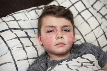
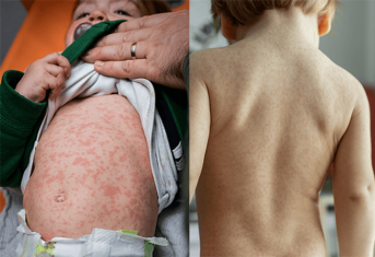
3. Skin rashes in children
There are three main situations in which a child may present with a more or less febrile rash:
- Rash with stereotyped course and good general condition: classic eruptive diseases and "eruptive syndromes" (viruses).
- Rash with non-stereotyped course +/- arthralgias, myalgias... but good general condition (virus).
- Rash immediately associated with altered general condition: bacterial infections, more rarely viral infections, inflammatory or systemic diseases.

There is no unambiguous relationship between the nature of the exanthem and the infectious agent responsible. Exanthems have been characterized on the basis of classic childhood diseases, but their cause can nowadays vary:
- Scarlatiniform exanthem: mainly bacterial infections, but also viral (Epstein-Barr virus [HBV]...).
- Morbilliform exanthema: mainly viral infections, but also bacterial, systemic lupus erythematosus, graft-versus-host disease.
- Roseoliform exanthema: viral infection (human herpesvirus type 6 [HHV6]), bacterial infection, Still's disease...

The list of main viruses responsible for exanthem in children has grown: enterovirus, HHV6, varicella-zoster virus, parvovirus B19, EBV, measles and rubella viruses, cytomegalovirus (CMV), rotavirus, echovirus, enterovirus...
This extension is due to the changes that have taken place between the 20th and 21st centuries, namely environmental modifications, zoonoses (child travellers), SARS-CoV-2, i.e., more generally, climatic and environmental changes, the progressive modification of endemic zones and seasonal epidemics.
The diagnostic approach must therefore be based, first and foremost, on an interview focusing on vaccinations, prodromal symptoms, the presence or absence of enanthema, accompanying signs, medication taken, epidemiology and the notion of travel. Clinical examination completes the diagnostic orientation: erosions suggest enterovirus, parvovirus, CMV or HIV, pharyngeal papules suggest HHV6 or HHV7, petechiae suggest EBV...
No unambiguous relationship between a characterized paraviral syndrome and the virus responsible...
Gianotti-Crosti syndrome (papular eruption of the face and extremities, lasting three to four weeks, with varying degrees of fever, in a context of good general health) has long been well characterized, and can occur with many viruses: HBV, EBV, CMV, HHV6, parvovirus B19, post-vaccination... The same picture can therefore be seen several times in the same child, linked to a different infectious agent.
Certain eruptions were described in the 20th century, such as eruptive pseudoangiomatosis: few angiomatous papules (less than ten lesions) with a vasoconstrictive halo, good general condition and spontaneous regression in one to two weeks. The first reported cases were linked to an Echovirus, but more recent publications report Coxsackies, adenovirus, CMV, EBV...
and for a given virus, several possible semiologies/evolutions
Parvovirus B19, for example, can produce a multitude of clinical pictures:
- Epidemic megalerythema.
- Non-specific rash, maculo-papular, vesiculo-papular.
- Mucosal lesions.
- Desquamation of extremities.
- Gianotti-Crosti syndrome.
- Purpura...
These data explain why, in the absence of serious clinical signs, a particular terrain or at-risk contacts, there is no need to investigate the viral origin of these symptoms, which will regress spontaneously.
From the 20th to the 21st century: new eruptions
Enterovirus eruptions have changed completely in recent years, with the classic hand-foot-and-mouth syndrome becoming less common, and the appearance of erythematous lesions on the face and limbs, vesicular lesions, perianal localization and erythema multiforme - all of which are becoming "classic".
Eruptive hypomelanosis is characterized by hypopigmented macules: the eruption is abrupt, taking three to four days to appear as well-limited macules, with no inflammatory phase or pruritus, on the arms, legs and extremities, with varying degrees of axillary adenopathy. Regression occurs spontaneously within three weeks. A viral etiology is strongly suspected, although the causative agent is not yet known.
Travel and climate change
Arboviroses such as Zika, dengue and chikungunya, once localized in the Caribbean, Central and South America, are now being seen in our climates, with symptoms combining fever, morbilliform rash, conjunctivitis and arthralgia to varying degrees.
(Based on a paper by Pr Christine Bodemer, Hôpital Necker-Enfants malades, Paris).
Articlewritten by Quotidien du Médecin in partnership with Ducray
Interested in our news?
Discover those of our other brands.
Want to read on?
This access is reserved for professionals, registered on Pierre Fabre For Med.
To access the full content, please register or log in if you already have an account.

